
Search
© 2024 khagapharmacy.com
NEUROSCIENCE
Contents
What is Neuroimaging: From Brain Structure to Facial PareidoliaIntroduction to Neuroimaging:Table of ContentsThe Need for Neuroimaging:Types of Neuroimaging Techniques:Applications of Neuroimaging:Advancements in Neuroimaging Technology:Limitations of Neuroimaging:Neuroimaging Unravels the Mysteries of Facial Pareidolia:FAQs about Neuroimaging: Conclusion:Leave a Reply Cancel reply
What is Neuroimaging: From Brain Structure to Facial Pareidolia
-
 Rahul Priydarss
Unlock the mysteries of facial pareidolia and delve into the fascinating world of neuroimaging with our comprehensive guide. From understanding the basics of neuroimaging techniques to exploring their diverse applications in research and clinical settings, discover how cutting-edge technology revolutionizes our understanding of the brain. Dive deep into advancements in neuroimaging technology, uncover the limitations, and gain valuable insights into the intricate workings of the human mind.
- Advertisement -
Introduction to Neuroimaging:
Neuroimaging is a field of study that involves various techniques to visualize the structure and function of the brain. It has revolutionized our understanding of the brain and its role in cognition, emotion, behavior, and neurological disorders. In this introduction, we’ll explore the basics of neuroimaging, its history, key techniques, and its significance in neuroscience and clinical practice.
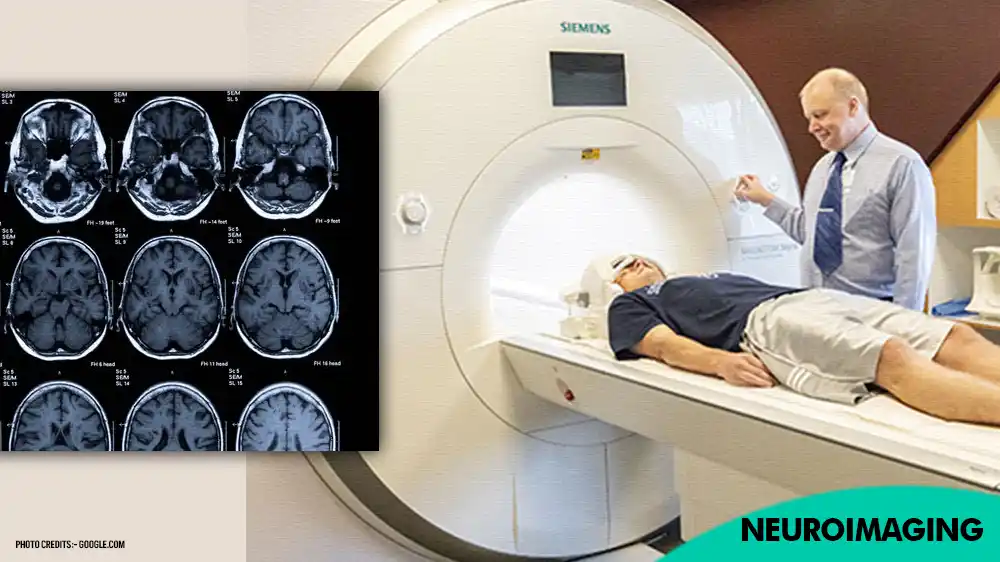
Table of Contents
The Need for Neuroimaging:
Neuroimaging plays a crucial role in understanding the complexities of the brain and diagnosing various neurological conditions. This section delves into the reasons why neuroimaging is essential.
Understanding Brain Anatomy and Function: Neuroimaging techniques allow researchers and clinicians to visualize the intricate structures and functions of the brain, aiding in unraveling its mysteries and complexities.
Diagnosis of Neurological Disorders: Neuroimaging helps in the diagnosis of neurological conditions such as strokes, tumors, traumatic brain injuries, neurodegenerative diseases (e.g., Alzheimer’s, Parkinson’s), epilepsy, and psychiatric disorders.
- Advertisement -
Treatment Planning and Monitoring: By providing detailed images of the brain, neuroimaging assists in treatment planning for neurological disorders, such as surgery for brain tumors or deep brain stimulation for movement disorders. It also aids in monitoring disease progression and treatment effectiveness.
Research and Advancement of Neuroscience: Neuroimaging techniques are invaluable tools for researchers studying brain development, cognitive processes, neural networks, and the effects of various interventions on brain function.
Personalized Medicine: In the era of precision medicine, neuroimaging contributes to personalized treatment approaches by providing insights into individual variations in brain structure and function, which can influence treatment outcomes.
Early Detection and Prevention: Early detection of neurological abnormalities through neuroimaging can enable timely intervention and preventive measures, potentially mitigating the progression of neurological diseases.
- Advertisement -
Enhancing Clinical Decision-Making: Neuroimaging findings contribute critical information to clinical decision-making, guiding physicians in choosing the most appropriate management strategies for patients with neurological conditions.
Types of Neuroimaging Techniques:
Neuroimaging encompasses various techniques that allow scientists and clinicians to visualize the structure, function, and activity of the brain. Here are some of the key types of neuroimaging techniques.
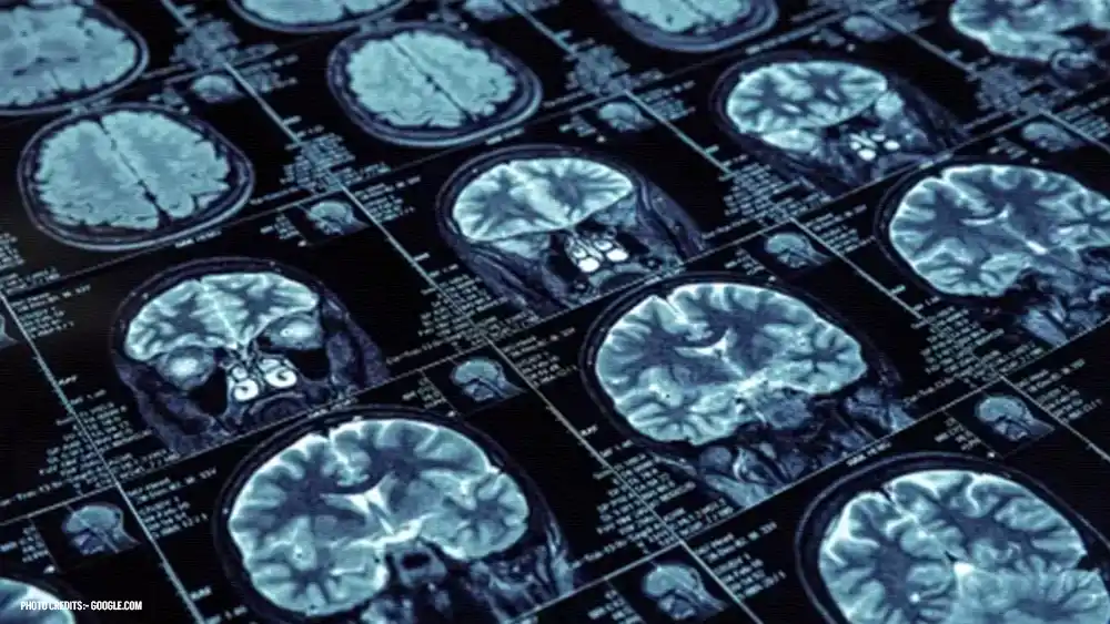
Structural Imaging: MRI (Magnetic Resonance Imaging): Uses magnetic fields and radio waves to generate detailed images of the brain’s structure, including soft tissues, such as the brain’s gray and white matter, and abnormalities like tumors or lesions.
CT (Computed Tomography): Involves X-ray technology to create cross-sectional images of the brain, useful for detecting acute conditions like bleeding or fractures.
Functional Imaging: fMRI (Functional Magnetic Resonance Imaging): Measures changes in blood flow and oxygenation levels in the brain, providing insights into brain activity during tasks or at rest.
- Advertisement -
- PET (Positron Emission Tomography): Uses radioactive tracers to detect changes in metabolic activity or neurotransmitter function in the brain, helpful in diagnosing conditions like Alzheimer’s disease or assessing tumor metabolism.
- SPECT (Single Photon Emission Computed Tomography): Similar to PET but uses different tracers to visualize brain activity, particularly useful in epilepsy localization and assessing blood flow abnormalities.
Diffusion Imaging: DTI (Diffusion Tensor Imaging): Maps the diffusion of water molecules in brain tissue, allowing visualization of white matter tracts and assessing structural connectivity between different brain regions.
Electrophysiological Imaging: EEG (Electroencephalography): Records electrical activity generated by the brain’s neurons through electrodes placed on the scalp, useful in diagnosing epilepsy and studying brain rhythms.
MEG (Magnetoencephalography): Measures the magnetic fields produced by neuronal activity, providing high temporal resolution and helping localize brain functions with precision.
Molecular Imaging: Molecular MRI: Involves the use of specialized contrast agents to visualize specific molecular targets in the brain, facilitating research on neurotransmitter systems and molecular processes underlying neurological disorders.
Applications of Neuroimaging:
Neuroimaging techniques have diverse applications in both research and clinical settings, offering insights into the structure, function, and activity of the brain. Here are some key applications.
- Advertisement -
Diagnosis and Monitoring of Neurological Disorders: Neuroimaging aids in the diagnosis and monitoring of various neurological conditions, including strokes, traumatic brain injuries, brain tumors, neurodegenerative diseases (e.g., Alzheimer’s, Parkinson’s), multiple sclerosis, epilepsy, and psychiatric disorders.
Brain Mapping and Localization of Functions: Functional neuroimaging techniques such as fMRI, PET, and MEG help map brain activity associated with specific tasks or sensory stimuli, aiding in the localization of cognitive functions, language processing, motor control, and sensory perception.
Treatment Planning and Guidance: Neuroimaging assists in treatment planning for neurological disorders, guiding surgical interventions (e.g., tumor resection, epilepsy surgery) and non-invasive treatments (e.g., transcranial magnetic stimulation, deep brain stimulation) by providing precise anatomical and functional information.
Understanding Brain Development and Plasticity: Longitudinal neuroimaging studies track brain development from infancy to adulthood, shedding light on the structural and functional changes that occur during maturation and how they relate to cognitive development, learning, and behavior. Neuroimaging also helps investigate brain plasticity and adaptation following injury or rehabilitation.
Research on Brain Disorders and Mechanisms: Neuroimaging enables researchers to study the underlying mechanisms of neurological disorders, such as aberrant neural circuits, neurotransmitter imbalances, and structural abnormalities, facilitating the development of novel therapeutic interventions and pharmacological treatments.
- Advertisement -
Personalized Medicine and Biomarker Discovery: Neuroimaging contributes to personalized medicine approaches by identifying biomarkers associated with disease progression, treatment response, and prognosis. It helps stratify patients based on neuroimaging signatures, leading to tailored treatment strategies and improved clinical outcomes.
Advancing Cognitive Neuroscience and Behavioral Studies: Neuroimaging techniques provide valuable tools for cognitive neuroscience research, allowing investigation of complex cognitive processes, emotion regulation, decision-making, and social behavior. They offer insights into the neural mechanisms underlying psychological disorders and individual differences in behavior and cognition.
Education and Public Awareness: Neuroimaging findings enhance public understanding of brain structure and function, fostering awareness of brain health, neuroscience research, and the impact of neurological disorders on individuals and society.
Advancements in Neuroimaging Technology:
Neuroimaging technology has witnessed significant advancements over the years, leading to improved spatial and temporal resolution, enhanced sensitivity, and expanded applications. Here are some notable advancements.
High-Field MRI: The development of high-field MRI systems, operating at 3 Tesla (3T) and beyond, has enabled higher spatial resolution and improved contrast in structural and functional imaging. Ultra-high-field MRI at 7 Tesla and higher offers unprecedented detail of brain anatomy and function, albeit with technical challenges related to image distortion and signal-to-noise ratio.
- Advertisement -
Multimodal Imaging Integration: Integrating multiple imaging modalities, such as combining structural MRI with functional MRI (fMRI), diffusion tensor imaging (DTI), and PET or SPECT, allows researchers and clinicians to obtain complementary information about brain structure, function, and metabolism in a single imaging session, enhancing diagnostic accuracy and research capabilities.
Functional Connectivity Mapping: Advancements in fMRI techniques, including resting-state fMRI and task-based fMRI paradigms, have facilitated the mapping of functional connectivity networks in the brain. These networks provide insights into the intrinsic organization of the brain and its alterations in health and disease.
Quantitative Imaging Biomarkers: Quantitative imaging techniques, such as diffusion kurtosis imaging (DKI), arterial spin labeling (ASL) perfusion imaging, and magnetic resonance spectroscopy (MRS), enable the measurement of physiological parameters and biochemical concentrations in the brain. These biomarkers offer valuable information for early detection, diagnosis, and monitoring of neurological disorders.
Real-Time Imaging and Feedback: Real-time functional neuroimaging techniques, such as real-time fMRI (rtfMRI) neurofeedback and functional near-infrared spectroscopy (fNIRS), allow individuals to observe and modulate their brain activity in real-time. These approaches hold promise for neurorehabilitation, cognitive enhancement, and treatment of neuropsychiatric conditions.
Machine Learning and AI Integration: Machine learning algorithms and artificial intelligence (AI) techniques are increasingly being integrated into neuroimaging analysis pipelines for automated image segmentation, feature extraction, pattern recognition, and predictive modeling. These tools accelerate data processing, improve diagnostic accuracy, and uncover complex relationships within large neuroimaging datasets.
- Advertisement -
Portable and Wearable Imaging Devices: Miniaturization of imaging technology has led to the development of portable and wearable neuroimaging devices, such as portable EEG systems, mobile MRI scanners, and wearable fNIRS headbands. These devices enable neuroimaging studies in diverse settings, including ambulatory monitoring, field research, and point-of-care diagnostics.
Ultrafast Imaging Techniques: Advancements in imaging sequences and reconstruction algorithms have facilitated ultrafast MRI techniques, such as echo-planar imaging (EPI), simultaneous multi-slice (SMS) imaging, and compressed sensing, allowing rapid acquisition of volumetric brain images with high temporal resolution. These techniques are particularly useful for real-time functional imaging and dynamic studies of brain function.
Limitations of Neuroimaging:
Despite its numerous benefits, neuroimaging also has several limitations that researchers and clinicians need to consider.
Spatial and Temporal Resolution: Most neuroimaging techniques have limitations in spatial and temporal resolution. While advancements have improved resolution over time, these limitations can affect the ability to detect small-scale brain structures or rapid changes in brain activity.
Cost and Accessibility: Neuroimaging equipment, such as MRI scanners and PET machines, can be expensive to acquire and maintain. Additionally, access to neuroimaging facilities may be limited in certain geographic regions, leading to disparities in healthcare and research opportunities.
- Advertisement -
Patient Cooperation and Comfort: Some neuroimaging procedures require patients to remain still for extended periods, which can be challenging for individuals with movement disorders, claustrophobia, or cognitive impairments. This can lead to suboptimal image quality and increased discomfort for patients.
Contrast and Sensitivity: Certain neuroimaging techniques may lack sufficient contrast or sensitivity to detect subtle changes in brain structure or function, particularly in cases where abnormalities are small or diffuse. This can pose challenges for accurate diagnosis and monitoring of neurological conditions.
Interpretation Challenges: Neuroimaging data interpretation requires specialized training and expertise. Without proper training, errors in image analysis or misinterpretation of findings may occur, leading to inaccurate diagnoses or conclusions.
Invasive Techniques: Some neuroimaging techniques, such as PET imaging with radioactive tracers or invasive electrophysiological methods, carry risks and ethical considerations associated with exposure to radiation or invasive procedures.
Biological Variability: There is considerable variability in brain structure and function among individuals, as well as within the same individual over time. This variability can complicate comparisons between different subjects or longitudinal studies and may require large sample sizes to draw meaningful conclusions.
- Advertisement -
Ethical and Privacy Concerns: Neuroimaging studies involving human participants raise ethical concerns regarding informed consent, privacy protection, and potential stigmatization based on imaging findings. Ensuring adherence to ethical guidelines and safeguarding participant confidentiality is essential in neuroimaging research.
Limitations in Studying Brain Dynamics: While neuroimaging techniques provide valuable insights into brain structure and static functional connectivity, they may have limitations in capturing dynamic changes in brain activity or interactions between brain regions over time.
Neuroimaging Unravels the Mysteries of Facial Pareidolia:
Imagine staring down at your breakfast and encountering the divine visage of Jesus Christ. This isn’t a scene from a religious text, but a real-life experience for Diana Duyser, who saw the Virgin Mary imprinted on a grilled cheese sandwich. Such occurrences, where we perceive faces in everyday objects, are attributed to a fascinating phenomenon called facial pareidolia.
For years, such sightings were relegated to the realm of religious fervor or extraterrestrial encounters. However, recent scientific breakthroughs are shedding light on the intricate neural processes underlying facial pareidolia. A groundbreaking study published in the Proceedings of the National Academy of Sciences (PNAS) delves into the “when” and “where” of this phenomenon within the brain.
The research, with its electroencephalogram (EEG) approach, aligns with a growing understanding of how our brains construct perception. It reveals a complex “dialogue” between different brain regions. Visual processing areas first encounter the stimuli, while memory regions attempt to fill in the gaps, drawing upon familiar patterns. Notably, the facial fusiform gyrus, specifically dedicated to facial recognition, plays a crucial role in the early stages of processing these perceived faces.
- Advertisement -
Our perception isn’t a passive recording of the world around us. It’s an active interpretation, shaped by anticipation and stored knowledge. When we see a familiar face, like Elvis Presley, our brain doesn’t meticulously analyze every detail. It leverages pre-existing representations, allowing for a swift and efficient recognition process. This is where things get interesting with pareidolia. Imagine spotting “Elvis” in a blob of ketchup. The “dialogue” between brain regions can go awry, leading to misinterpretations. We end up seeing a face where none exists.
This tendency to perceive faces, particularly those we’re familiar with, can be explained by the way we store information in memory. Faces hold a special significance, and our brains seem pre-wired to seek them out. Studies by Dr. Susana Martínez-Conde, a neurologist, demonstrate this through eye-tracking experiments. Participants demonstrably spend more time fixated on faces compared to other objects. From an evolutionary standpoint, this makes perfect sense. As social creatures, recognizing faces of friends, family, and potential threats is crucial for survival.
Interestingly, pareidolia reveals more about the perceiver than the perceived. Our inherent biases shape these optical illusions. A study published in PNAS found a significant bias towards perceiving masculine faces. Over 80% of participants assigned a male gender to these illusory faces. This tendency extends beyond toast or potatoes; we seem to see men’s faces everywhere.
This phenomenon goes beyond just faces. We might hear our name being called in a random noise, or decipher hidden messages in audio recordings. Our brains are wired to find meaning, even in disorganized information. Perception is less about a perfect reconstruction of reality and more about a constructed simulation.
However, our brains also possess a corrective mechanism. After the initial perception of a face, we usually re-evaluate the situation. We understand that the burnt toast isn’t Jesus Christ (unless fueled by strong religious beliefs or mental illness). This process of reevaluation can be disrupted in certain neurodegenerative diseases, particularly Parkinson’s. Studies indicate that a significant portion of Parkinson’s patients experience facial pareidolia, with the perceived faces becoming integrated into their reality.
- Advertisement -
Understanding facial pareidolia offers valuable insights into both how we perceive the world and the workings of the brain. By pinpointing the brain areas involved, this research not only confirms that the toast isn’t a deity but also helps us gain a deeper understanding of ourselves and the fascinating ways in which our brains construct our reality. As Dr. Martínez-Horta aptly concludes, “Pareidolia tells us very well the fact that we have no control over what we are seeing is an example of how we often perceive the world.
FAQs about Neuroimaging:
A1: Yes, neuroimaging techniques like MRI and CT scans are considered safe when performed by trained professionals.
A2: While neuroimaging can provide valuable insights, mental illness diagnosis typically involves a comprehensive evaluation by healthcare professionals.
A3: The duration varies depending on the type of scan and the specific protocol but typically ranges from 15 minutes to an hour.
A4: Neuroimaging procedures are generally safe, but some individuals may experience claustrophobia in MRI machines or allergic reactions to contrast agents.
- Advertisement -
A5: In many cases, neuroimaging procedures are covered by health insurance, but coverage may vary depending on the type of scan and individual insurance plans.
-Please remember, to always consult with healthcare professionals or Doctors for personalized advice related to medical conditions.
Conclusion:
Here is Neuroimaging, a cornerstone of modern neuroscience, offers unprecedented insights into the structure, function, and dynamics of the human brain. From unraveling the mysteries of facial pareidolia to diagnosing neurological disorders and guiding personalized treatment approaches, neuroimaging plays a pivotal role in advancing both research and clinical practice.